- 3 February 2021
Occlusion (blockage) of a retinal vein is a common cause of sudden painless reduction in vision in older people.
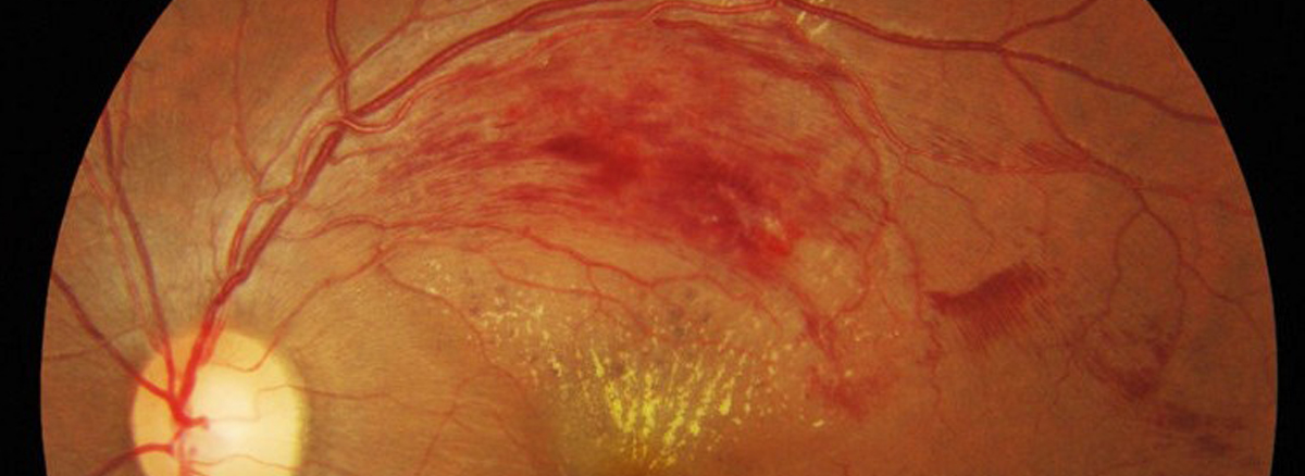
The retina is the thin membrane that lines the inner surface of the back of your eye. Its function is similar to that of the film in a camera. Blockage of one of the veins draining blood out of the eye causes blood and other fluids to leak into the retina, causing bruising and swelling as well as lack of oxygen. This interferes with the light receptor cells and reduces vision.
The condition is uncommon under the age of 60 but becomes more frequent in later life.
There are two main types of retinal vein occlusion:
- Branch retinal vein occlusions are due to blockage of one of the four retinal veins, each of which drains about a quarter of the retina
- Central retinal vein occlusion is due to blockage of the main retinal vein, which drains blood from the whole retina
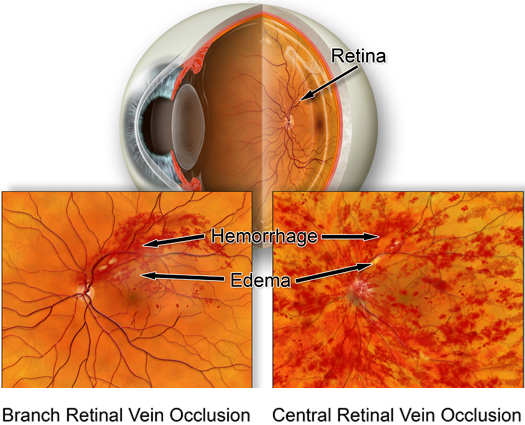
In general, visual loss is more severe if the central retinal vein is blocked.
What causes retinal vein occlusion?
A blockage forms in the vein, usually due to a blood clot, and obstructs the blood flow. The exact cause is unknown, but several conditions make the condition more likely. These include:
- High blood pressure
- High cholesterol
- Glaucoma
- Diabetes
- Smoking
- Certain rare blood disorders
Prevention and treatment
It is essential to identify and treat any risk factors to minimise the risk to the other eye and prevent a further vein occlusion in the affected eye although, in a small number of cases, no risk factors can be found, with the cause being unknown. Treatment of any risk factors detected reduces the risk of a further vein occlusion occurring in either eye and may also help to reduce the risk of other blood vessel blockages, such as may happen in a stroke (affecting the brain) or a heart attack or, in those with rare blood disorders, a blocked vein in the leg (deep vein thrombosis) or lung (pulmonary embolism).
Persistent bruising and swelling at the centre of the retina (the macula) is the main cause of permanent loss of central vision. The swelling is caused by damaged blood vessels which leak fluid. Different medicines such as anti- vascular endothelial growth factor (anti-VEGF) medicines or steroids may be helpful in reducing this leakage. These medicines are given by injection into the eye and the injections often need to be repeated as the effect of the medicine wears off. Laser treatment is sometimes helpful in restoring some central vision in branch retinal vein occlusions.
About 20% of patients with retinal vein occlusions develop abnormal blood vessels growing on the iris at the front of the eye or on the retina. These abnormal blood vessels can bleed or cause a marked pressure rise in the eye leading to further loss of vision. This can normally be prevented by laser treatment to the retina, which is most effective if applied before vision is lost. For this reason, patients with central retinal vein occlusions are normally checked every four to six weeks for six months but branch retinal vein occlusions can be checked less often as the risk is much less.
The following three tests, which provide digital images of the retina and its blood circulation, are frequently recommended for patients with retinal vein occlusion to help monitor the condition and decide the most appropriate treatment:
- Retinal photography
- Fluorescein angiography
- Optical coherence tomography
Most patients are discharged after four years as recurrence or deterioration is unlikely after this.